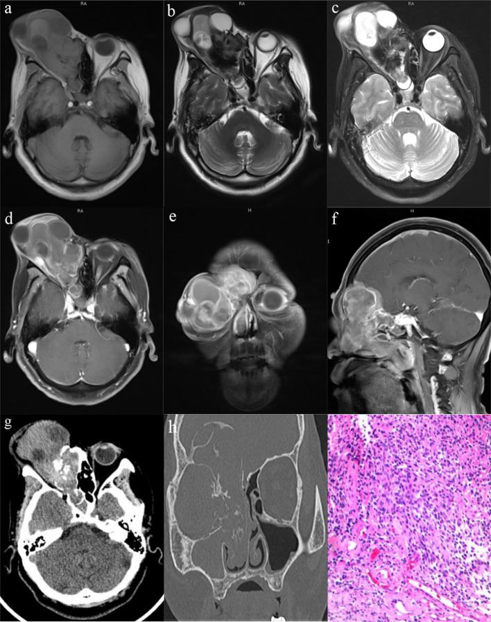
Fig. 1.
Pre-operative contrast-enhanced MRI and CT scan of the brain and paranasal sinuses and the histopathological examination with hematoxylin and eosin stained. a–c Non-contrast MRI revealed heterogeneous isointense signal of the mass on T1-weighted sequences and hypointense to hyperintense signal on T2-weighted sequences and fat attenuated sequences, with unclear boundaries and large invasion range. d–f Contrast-enhanced MRI showed dumbbell shaped mass with intense heterogeneous post contrast enhancement in the right superior nasal cavity with extensions into the right orbit, anterior cranial fossa and paranasal sinuse. Peritumoral cysts are also noted at the tumor brain interface. g, h CT scan of the brain and paranasal sinuses revealed that there was erosion of the right paranasal sinuses wall, the cribriform plate, and the right medial orbital wall with significant bony destruction and radial periosteal reaction. The ‘waist’ of the dumbbell shaped mass is at the cribriform plate. i Hematoxylin and eosin stained slide (× 200)—showing olfactory neuroblastoma which is having a high nuclear: cytoplasmic ratio, round hyperchromatic nuclei with inconspicuous nucleoli, scanty cytoplasm, and a richly intercellular vascularized fibromyxoid stroma

