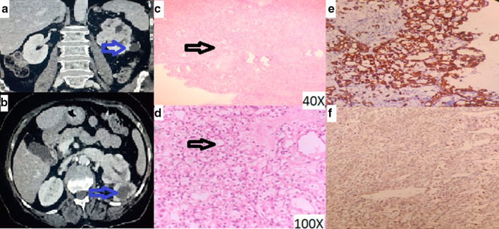
Fig. 2.
a Enhancing left renal mass b Enhancing left renal mass with left renal vein invasion c 40X image showing tumour cells arranged in diffuse sheets d 100X Image showing large tumor cells with clear and vacuolated cytoplasm, round to oval nucleus and prominent nucleolus e Tumor cells positive for Vimentin on IHC f Tumor cells showing positivity for RCC antigen on IHC

