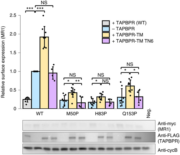
Fig. 4 ∣. Indirect assessment of TAPBPR/MR1 interactions in cells.
Wild-type or proline mutants of human MR1 (x axis), with an extracellular c-myc tag, were expressed in Expi293F cells without or with FLAG-tagged TAPBPR, TAPBPR-TM or TAPBPR-TM TN6. Top, surface levels of MR1, measured by flow cytometry (representative data and gating strategy shown in Extended Data Fig. 7). Statistical significance was assessed using one-way ANOVA: NS, not significant; *P < 0.05, **P < 0.01, ***P < 0.001. A full list of P values is noted in the Methods. Bottom, western blots show total protein expression. Anti-cyclophilin B is a loading control. Neg., vector-only transfected cells. Data are mean ± s.d. from the following number of independent experiments: N = 4, MR1 H83P + TAPBPR-TM TN6; N = 5, MR1 Q153P + TAPBPR-TM TN6, MR1 M50P + TAPBPR-TM TN6, MR1 + TAPBPR-TM TN6; N = 7, MR1 + WT TAPBPR, MR1 H83P-TAPBPR; N = 8, MR1 M50P-TAPBPR, MR1 H83P + TAPBPR-TM, MR1 Q153P-TAPBPR; N = 9, MR1 -TAPBPR, MR1 + TAPBPR-TM, MR1 M50P + TAPBPR-TM, MR1 Q153P + TAPBPR-TM.

