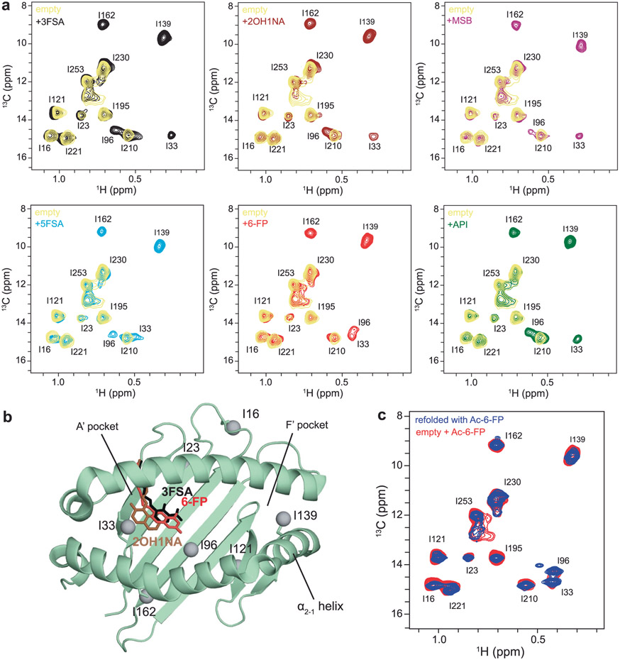
Extended Data Fig. 3 ∣. Chemical shift profiles of Ile 13Cδ1 labeled hpMR1 loaded with different ligands.
a, 2D 1H-13C methyl HMQC spectra of 10 μM Ile 13Cδ1-labeled hpMR1 with natural isotopic abundance bβ2m in the absence (empty, yellow) and presence of 2 mM ligand recorded at a 1H field of 600 MHz at 25 °C. All samples contain 0.5% DMSO-d6. b, Ile δ1 methyl groups (grey spheres) mapped onto the MR1 groove in complex with 6-FP (PDB ID 4GUP, red), 3FSA (PDB ID 5U6Q, black, TCR removed), or 2OH1NA (PDB ID 5U16, brown, TCR removed). c, Overlay of 2D 1H-13C methyl HMQC spectra of Ile δ1 labeled hpMR1 with natural isotopic abundance bβ2m. Samples were derived from in vitro refolding of the Ac-6-FP/Ile labeled hpMR1/bβ2m complex (blue) or from titration of empty Ile δ1 labeled hpMR1/bβ2m with 2 mM Ac-6-FP (red). Refolded and titrated spectra were recorded at 25 °C at a 1H field of 800 MHz and 600 MHz, respectively.

