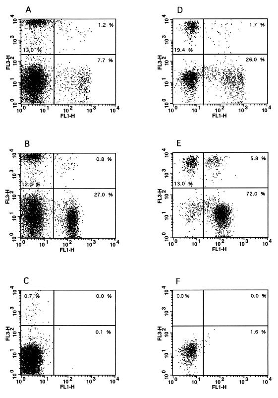
FIG. 3.
Two-color flow cytometric analysis of bovine blood and milk lymphocytes. Purified lymphocytes were isolated from the blood (A to C) and milk (D to F) of cows with mastitis, and two-color flow cytometric analysis was performed as described in Materials and Methods. (A and D) Staining of blood (A) and milk (D) lymphocytes with antibody GD3.8 (FL3, specific for γδ T cells) versus antibody CC58 (FL1, specific for bovine CD8). (B and E) Staining of blood (B) and milk (E) lymphocytes with antibody GD3.8 (FL3, specific for γδ T cells) versus antibody CC42 (FL1, specific for bovine CD2). (C and F) Control staining levels with secondary antibodies only in blood (C) and milk (F) samples. The data are representative of at least five independent experiments.

