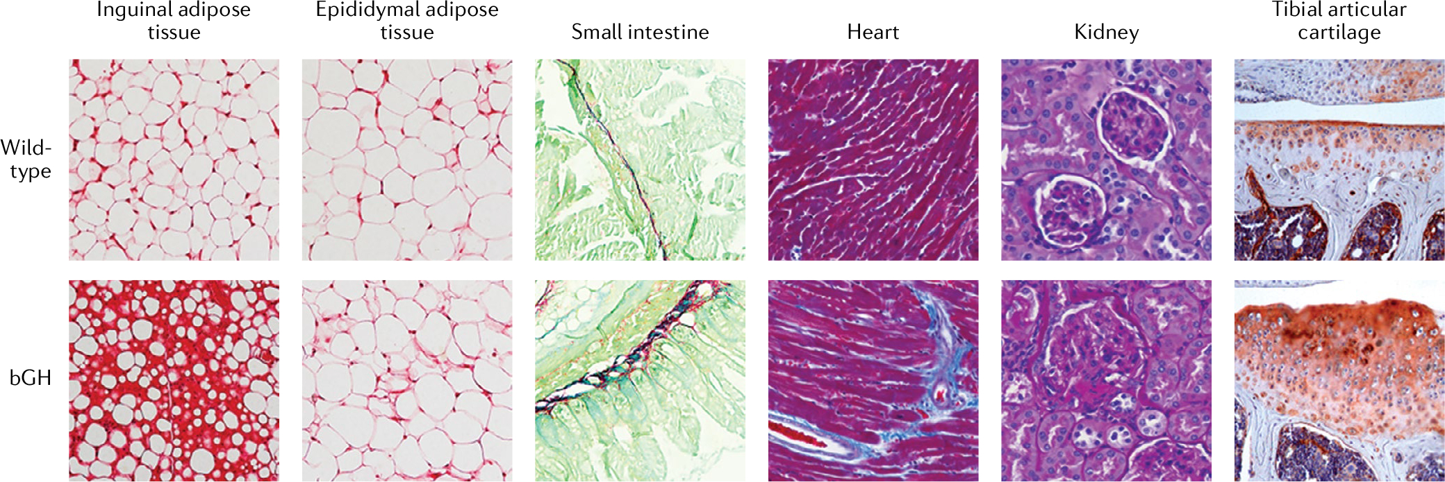
Fig. 1 |. GH overexpression in mice induces tissue fibrosis.

Histology of assorted tissues in aged male mice (10–13 months of age) overexpressing bovine growth hormone (bGH) and wild-type control mice. White adipose tissue from inguinal or epididymal adipose depots was stained with Sirius red (a non-specific red collagen stain), Swiss rolls of the small intestine were stained using Sirius Red and Fast Green (which stains non-collagenous protein), heart was stained with Masson’s trichrome (which stains connective tissue blue) and kidney sections were stained with periodic acid Schiff stain (which stains connective tissue a purple–magenta colour). The tibial articular cartilage images were generated via immunostaining with a type X collagen (a bone-specific collagen)-specific antibody (orange–red colour). In bGH mice, the increased GH activity results in increased fibrosis in the tissues shown. The histological images of tibial articular cartilage are courtesy of S. Zhu, Ohio University.
