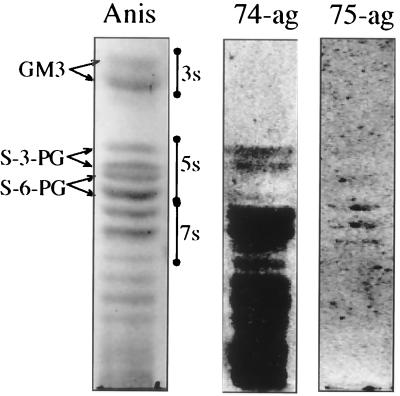
FIG. 1.
Gangliosides of human leukocytes separated on silica gel TLC plates and visualized by anisaldehyde (Anis), 35S-labeled H. pylori CCUG 17874 grown on agar (74-ag), or 35S-labeled H. pylori CCUG 17875 grown on agar (75-ag). 3s, 5s, and 7s indicate migration regions for three-, five-, and seven-sugar-containing monosialogangliosides, respectively. The TLC plates were developed in chloroform–methanol–0.25% KCl in water (50:40:10, by volume).

