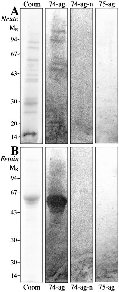
FIG. 3.
SDS-PAGE of human neutrophil protein extract from fresh membranes (Neutr, panel A) and of calf fetuin (Fetuin, panel B) on a 12.5% polyacrylamide homogeneous gel stained with Coomassie brilliant blue (Coom) and the corresponding autoradiograms after binding of 35S-labeled H. pylori CCUG 17874 on PVDF membrane blot (74-ag), strain CCUG 17874 on neuraminidase-treated PVDF membrane blot (74-ag-n), and strain CCUG 17875 on PVDF membrane blot (75-ag). The numbers on the left denote apparent relative molecular masses in kilodaltons.

