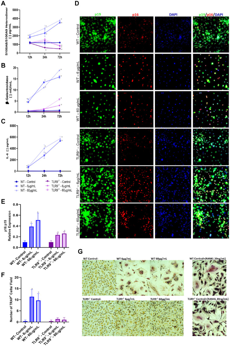
Figure 4.
Ex vivo evaluation of inflammaging and senescence markers in bone marrow–derived macrophages (BMDMs) isolated from wild-type (WT) and TLR9–/– that were stimulated with varying concentrations of TLR9 ligand (ODN 1668) (6 µg/mL and 60 µg/mL) or left unstimulated over different time points (12 h, 24 h, and 72 h). Enzyme-linked immunosorbent assay was used to measure (A) S100A8/A9 heterodimer (calprotectin) accumulation in BMDM cell lysate and (B, C) levels of β-galactosidase and interleukin-6 (Il-6) in BMDM cell supernatant, respectively. (D, E) Immunofluorescence (IF) for p16INK4a, p19ARF, and DAPI was performed at the 72-h time point. (D) Representative IF images taken at 40× magnification. (E) Balance of the p16INK4a/p19ARF expression in relation to DAPI. (F, G) Evaluation of osteoclast differentiation (purple/TRAP+ cells) over 6-d stimulation with ODN. Treatment with RANKL (20 ng/mL) was used as positive control for osteoclast differentiation. (F) Number of osteoclasts (TRAP+ cells) per field at 40× magnification. (G) Representative images for TRAP staining. (*) indicates significant difference in relation to BMDMs isolated from WT mice not stimulated with ODN (negative control). One-way analysis of variance with post hoc Tukey’s test was used at the significance level of 5%. TRAP, tartrate-resistant acid phosphate.

