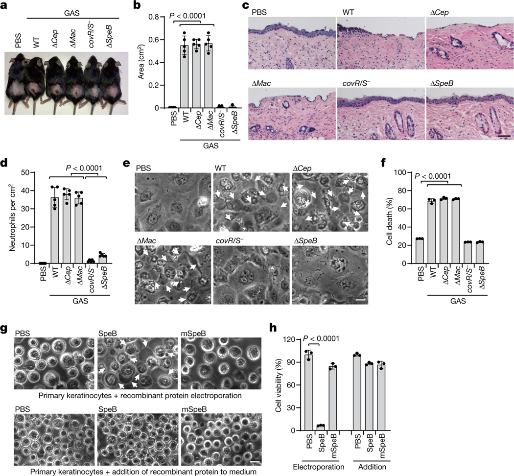
Fig. 1 |. The GAS virulence factor SpeB triggers lytic death of skin epithelial cells.
a–d, Mice were infected with GAS M1T1 strain 5448 (WT) or its isogenic mutant strains (ΔcepA, Δmac, covR/S− and ΔspeB). a, Representative image of skin lesions of mice challenged with indicated GAS variants. b, Quantification of skin lesion size. c, Histopathology of skin biopsies analysed by haematoxylin and eosin (H&E) staining. Scale bar, 100 μm. d, Quantification of neutrophil infiltration at skin lesion site. e, f, Primary mouse keratinocytes infected with indicated GAS strains for 2.5 h or not infected (PBS) were analysed by phase-contrast microscopy (e) and assayed for LDH release (f). g, h, Equal amounts of recombinant WT SpeB or the protease activity-deficient mutant mSpeB were electroporated into primary keratinocytes for 1 h, followed by observation of cell morphology by phase-contrast microscopy (g) and cell viability assessment by CellTiter-Glo luminescent assay (h). Keratinocytes displayed a rounded shape as they were resuspended before treatments. Arrows indicate pyroptotic cells. Scale bar, 10 μm. Data in b, d, are mean ± s.d. (n = 5 mice per group); data in f, h, are mean ± s.d. of triplicate wells. b, d, f, One-way analysis of variance (ANOVA). h, Two-tailed Student’s t-test. Data are representative of at least three independent experiments.

