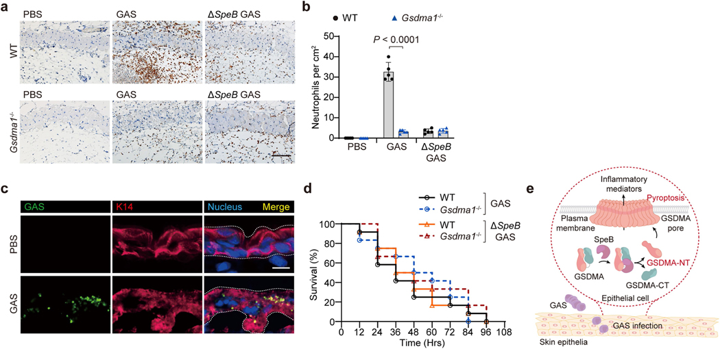
Extended Data Fig. 11 |. Gsdma1 deficiency affects host immune responses against subcutaneous but not intraperitoneal infection of GAS WT.
a, WT and Gsdma1−/− mice were subcutaneously infected or not with GAS isolate M1T1 strain 5448 or its isogenic mutant strain (ΔspeB variant). IHC analysis of neutrophil infiltration at infection site on day 1. Scale bar: 100 μm. b, Quantification of neutrophil infiltration at infection site. c, Cutaneous sections from Gsdma1−/− mice infected or not for 18 h with FITC-labelled GAS WT were subjected to immunofluorescence staining with anti-keratin 14. Nucleus was stained with DAPI. Dashed lines delineate the boundaries of epidermis. Scale bar: 10 μm. d, Survival rate of mice intraperitoneally administrated with the indicated GAS (n = 12 mice per group). e, Model of SpeB-triggered GSDMA activation and subsequent pyroptosis of skin epithelial cells during GAS infection. b, show mean ± s.d. (n = 5 mice per group); Two-tailed Student’s t-test. Data are representative of at least three independent experiments.

