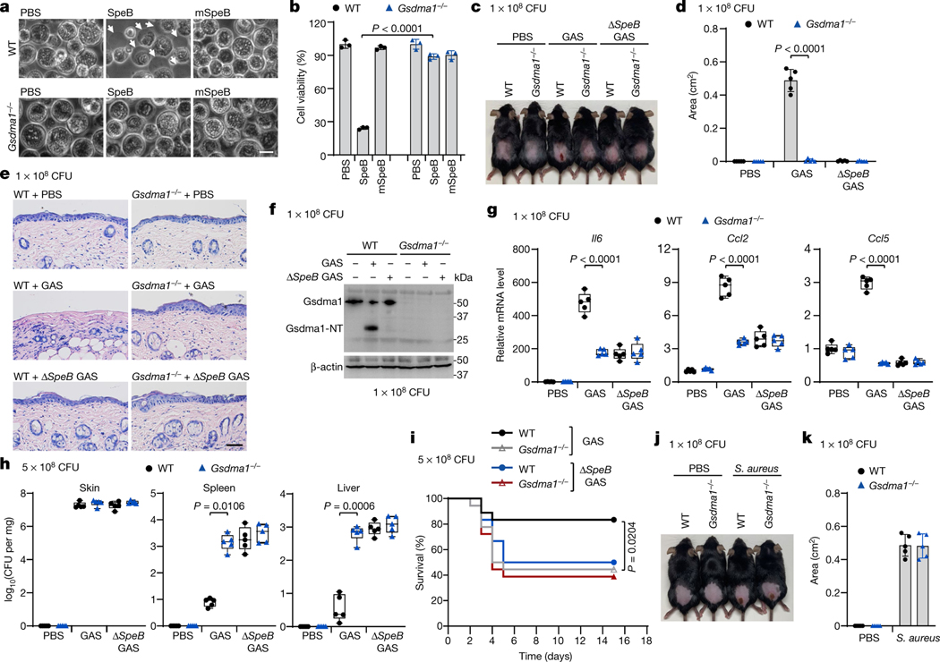
Fig. 4 |. Gsdma1 deficiency blunts host anti-GAS immunity.
a, b, Primary keratinocytes isolated from WT or Gsdma1−/− mice were electroporated with recombinant SpeB. Cell morphology was observed by phase-contrast microscopy (a) and cell viability was assessed by CellTiter-Glo luminescent assay (b). Arrows indicate pyroptotic cells. Scale bar, 10 μm. c–i, WT and Gsdma1−/− mice were infected with GAS M1T1 strain 5448, ΔspeB GAS or PBS control. c, Representative image of skin lesions of mice challenged for 1 day. d, Quantification of skin lesion size. e, Histopathology of skin biopsies analysed by H&E staining. Scale bar, 100 μm. f, Skin biopsies from WT or Gsdma1−/− mice infected with WT or ΔspeB mutant GAS were analysed by western blot. g, The induction of Il6, Ccl2 and Ccl5 expression in skin biopsy was examined. h, Bacteria load measured from skin lesions, spleens and livers of mice infected with GAS. i, Survival rate of mice challenged with the indicated GAS variants (n = 18 mice per group). j, k, WT and Gsdma1−/− mice were subcutaneously injected with S. aureus or PBS for 3 days. j, Representative image of skin lesions of mice. k, Quantification of skin lesion size. b, Data are mean ± s.d. of triplicate wells. d, k, Data are mean ± s.d. (n = 5 mice per group). g, h, Box plots show all data points, whiskers extend from minimum to maximum values (n = 5 mice per group), and the centre line, upper limit and lower limit of the box denote median, 25th and 75th percentiles, respectively. b, d, g, h, Two-tailed Student’s t-test. i, Mantel–Cox log-rank test. Data are representative of at least three independent experiments. CFU, colony-forming units. For gel source data, see Supplementary Fig. 1.

