Figure 2.

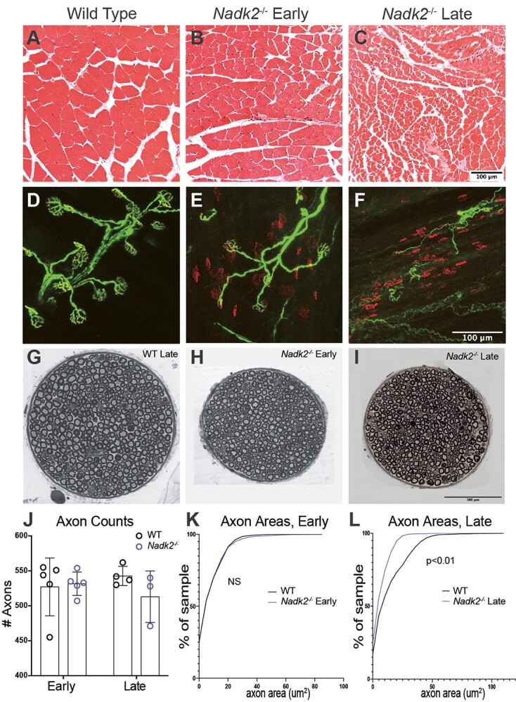
Nadk2 −/− muscle and nerve histology. (A–C) Cross-sections of the gastrocnemius muscle stained with H&E in wild type (A), 5-week-old Nadk2−/− (B), and 11-week-old Nadk2−/− mice (C). Severe muscle atrophy was present at 11 weeks. (D–F) Neuromuscular junctions in the plantaris muscle in wild type (D), 5-week-old Nadk2−/− (E), and 9-week-old Nadk2−/− mice (F). Neurofilament and SV2 label the nerve (green) and a-Bungarotoxin labels the postsynaptic acetylcholine receptors (red). Many denervated NMJs are observed at both 5 and 11 weeks of age. (G–I) Cross-sections of the motor branch of the femoral nerve in 11-week-old wild-type (G), 5-week-old Nadk2−/− (H), and 11-week-old Nadk2−/− mice (I). (J) The number of myelinated axons in the femoral motor nerve was not reduced at either age in Nadk2−/− mice. (K–L) The distribution of axon cross-sectional diameters was not different than control at 5 weeks of age (K), but axon size did not increase with age and axons in Nadk2−/− mice were smaller than wild type at 11 weeks of age. Scale bar in (C) is 100 μm for (A–C), in (F) is 100 μm for (D–F), and in (I) is 500 μm for (G–I). Significance tested by the Student t-test (J), or Kologmorov–Smirnoff and Mann–Whitney U tests (K, L).
