
Figure 1. Hypothesized signaling in the BLA and CeA relevant to alcohol use disorder.
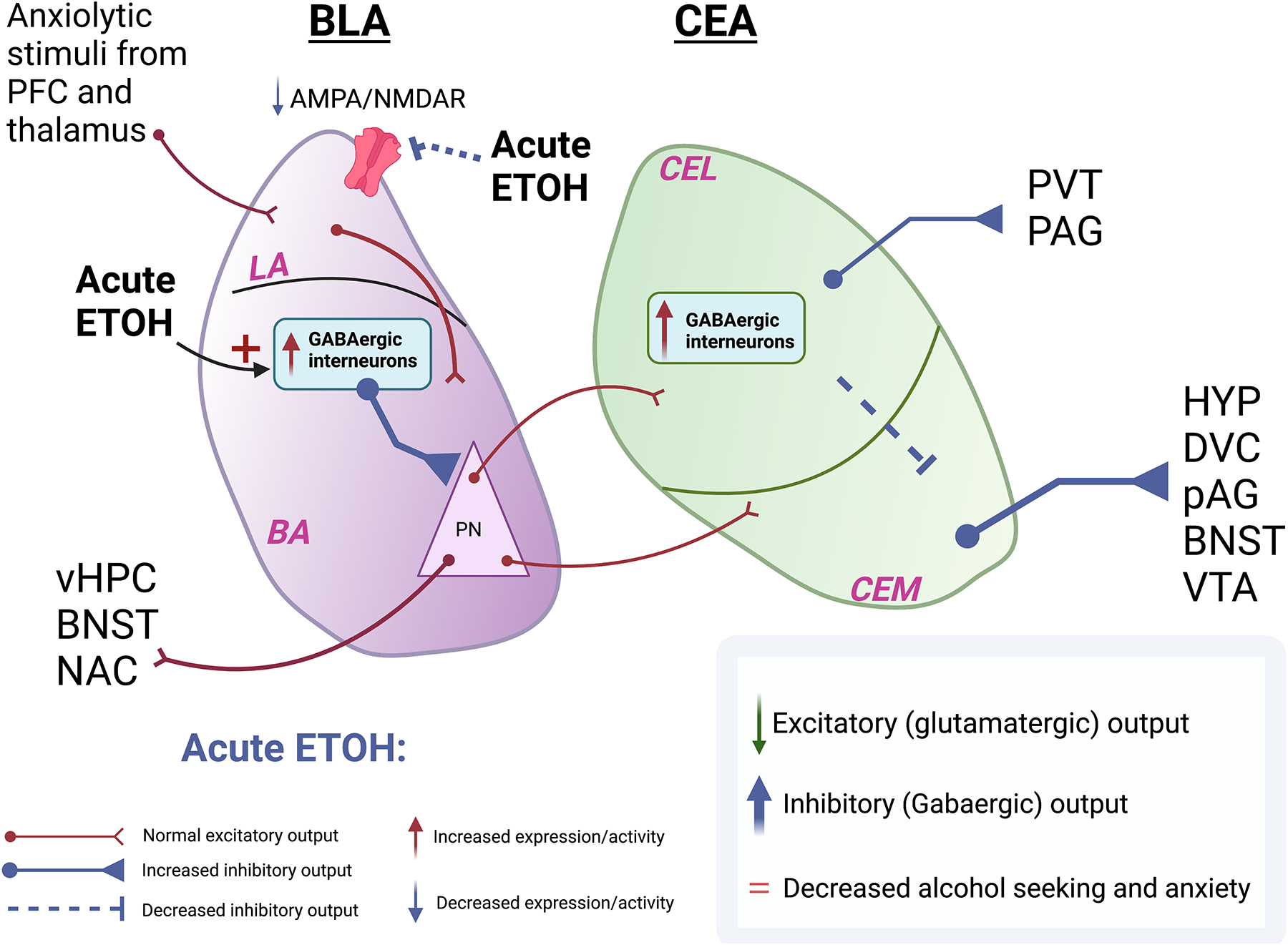
A) Following acute alcohol exposure there is a transient increase in inhibitory signaling onto excitatory pyramidal neurons in the BLA, leading to dampening of glutamatergic transmission. The CeA, in turn, receives decreased innervation from the BLA, leading to increased inhibitory output from the CeA. B) Following chronic ethanol exposure there is a decrease in GABAergic transmission from the interneurons in the BLA onto the pyramidal excitatory neurons of the BLA. There is also increased glutamatergic transmission within the BLA resulting from an increase in the AMPA/NMDAR ratio. Despite a decrease in CEL interneuron signaling, glutamatergic neurons in CeA receive greater input from PKCd+ interneurons from the CeM. The inhibitory output from the CeA is therefore dampened leading to an increase in alcohol seeking and anxiety.
Abbreviations: PFC: Prefrontal Cortex; BLA: Basolateral Amygdala; CEA: Central Amygdala; CEL: Centrolateral Amygdala; CEM: Centromedial Amygdala PV+: GABAergic parvalbumin-expressing interneurons; PKCd+: GABAergic protein kinase C delta-expressing interneurons; PN: Pyramidal Neurons; vHPC: Ventral hippocampus; BNST: Bed nucleus of stria terminalis; NAC: Nucleus accumbens; HYP: Hypothalamus; DVC: Dorsal vagal complex; VTA: Ventral tegmental area; PAG: Periaqueductal grey; PVT: Paraventricular nucleus of the thalamus