
Figure 2.
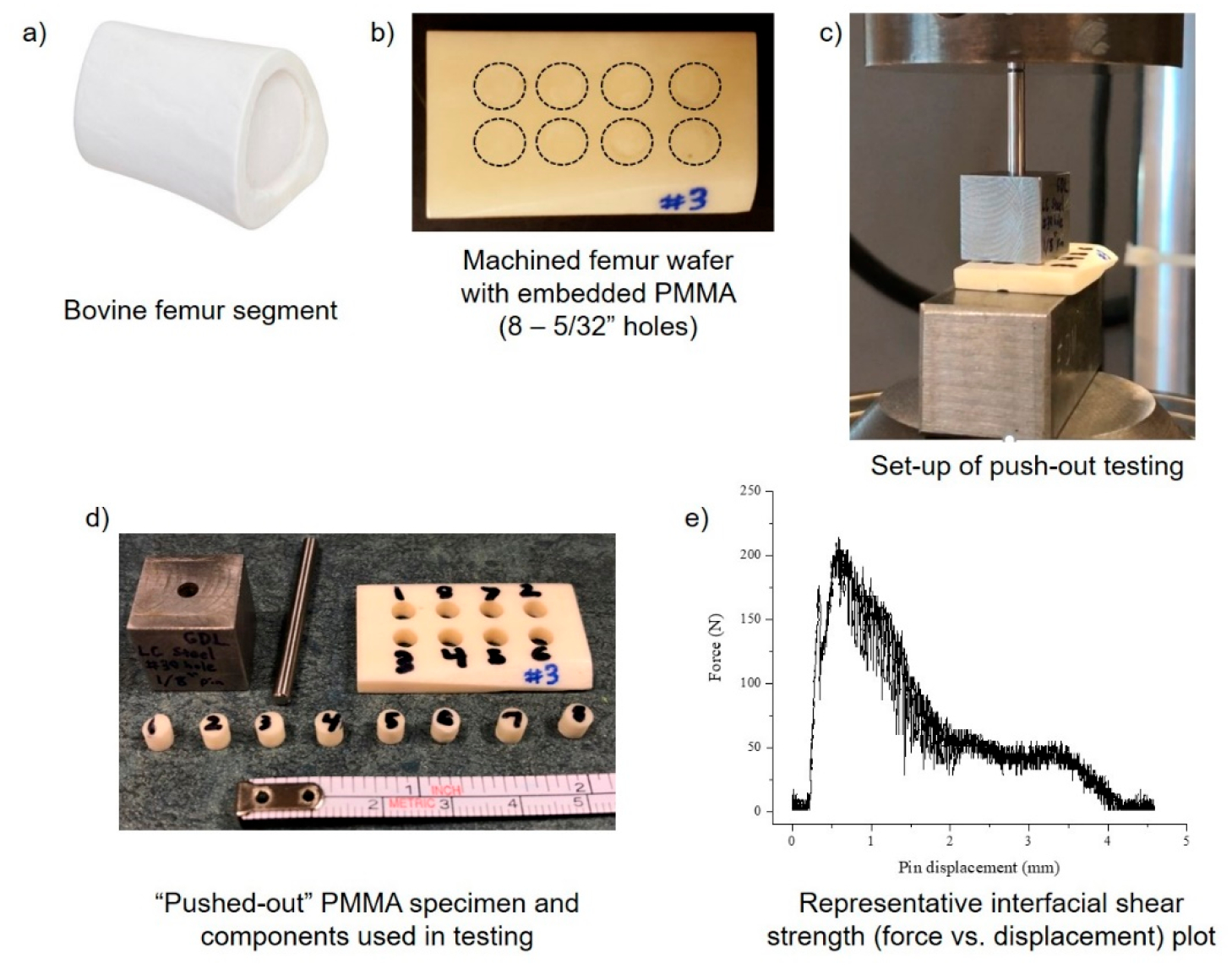
Setup of ex vivo PMMA bone cement push-out testing model using bovine femur tissue. Small, uniform wafers of bovine femur were machined from a cleaned/bleached femur segment (a) with eight 5/32 in. holes spaced 0.3 in. apart (b). Holes in the femur wafer were packed with PMMA materials, and a 1/8 in. steel pin was used to push embedded PMMA out of the femur wafer under compressive force at a loading rate of 20 mm/min (c). “Pushed-out” PMMA specimen from femur wafer (d). Representative raw force versus displacement plot of interfacial shear strength of PMMA pushed-out of a single 5/32 in. hole (e). Images were taken by the authors.