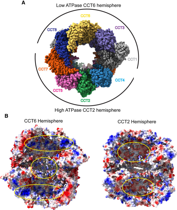
Figure 2. Functional differences between the CCT hemispheres.
(A) Open form of human CCT (PDB 6QB8) showing the hemispheres centered around CCT2 and CCT6 with high ATPase activity in the CCT2 hemisphere and low ATPase activity in the CCT6 hemisphere. (B) Electrostatic surface views of the interior of the closed form of human CCT (PDB 7NVN) with the CCT6 hemisphere on the left and the CCT2 hemisphere on the right. (PDB 7NVN, positive charge = blue, neutral = white, and negative charge = red.) For the CCT6 hemisphere, the CCT2, 4, 5 and 7 subunits have been removed to reveal the interior of the folding chambers. Positively charged patches in the upper and lower chambers and between the CCT rings are highlighted by yellow ovals. For the CCT2 hemisphere, the CCT1, 3, 6 and 8 subunits have been removed to view the interior of the folding chambers. Negatively charged patches in the upper and lower chambers are highlighted by yellow ovals.

