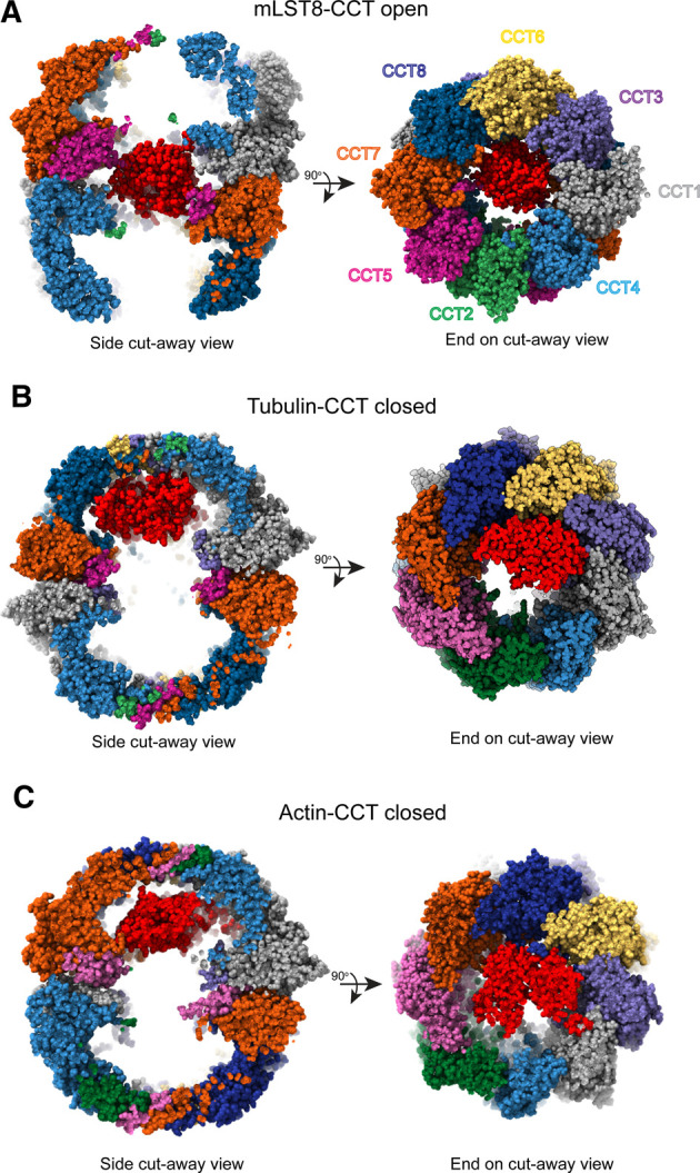
Figure 3. Substrate binding positions within CCT.

(A) Side and end-on cut away views of mLST8 (PDB 4JT6) sitting between the rings in the open form of human CCT (PDB 6QB8) [12]. (B) Side and end-on cut away views of β-tubulin in the folding cavity of closed CCT (PDB 7NVN) [32], highlighting the position of tubulin bound in the apical domains of the CCT6 hemisphere. (C) Side and end-on cut away views of actin in the folding cavity of closed CCT (PDB 7NVM) [32], showing actin spanning the cavity to interact with both hemispheres. Substrates are shown in red.
