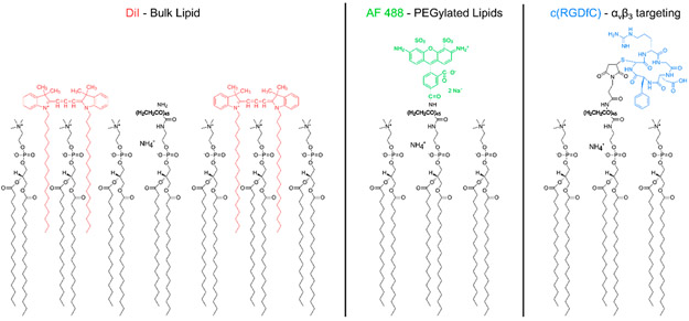
Figure 1.
Fluorophores and targeting peptides in relation to the lipid droplet shell. DiI (left) was used as a marker for DSPC, the primary or “bulk” lipid present on the droplet shell. Alexa Fluor (AF) 488 (center) was used as a marker for PEGylated lipids. For the binding assay experiments, a cyclic RGD peptide (right) was covalently linked to the terminal maleimide of the PEGylated lipids. The orientation of the markers and targeting peptide with respect to the lipid shell is shown. The 2000 molecular weight PEG chain is bolded. The shell composition is not to scale; the true ratio of DSPC to PEGylated lipids was 90:10 mol % for all droplets.

