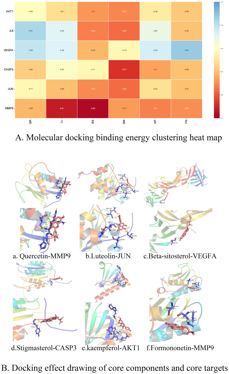
Fig 5. Molecular docking diagram.
Note: A. Heat map of molecular docking binding energy. The smaller the value of molecular docking binding energy is, the docking is stable, and the color changes with the value of binding energy. Quercetin(QR), luteolin(LL), beta-sitosterol (BS), Stigmasterol (SM), kaempferol (KP), formononetin (FN). B. Docking effect drawing of core components and core targets.

