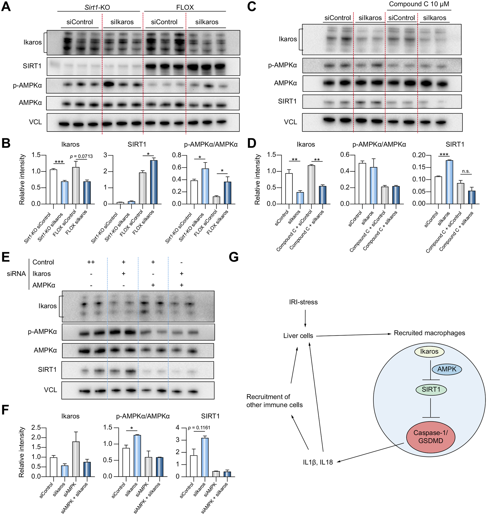
Fig. 7. Negative regulation of SIRT1 by Ikaros is AMPK dependent.

(A) BMMs from mSirt1-KO and FLOX mice were transfected with siControl or siIkaros RNAs. Lysates were probed by western blots for Ikaros, SIRT1, p-AMPKα, AMPKα and VCL as a loading control. (B) Relative intensity ratios with VCL normalization (n = 3). (C) wild-type BMMs transfected with siControl or siIkaros RNAs were pre-treated with Compound C (10 μM, 2 h). Lysates were probed by western blots for Ikaros, p-AMPKα, AMPKα, SIRT1 and VCL expression. (D) Relative intensity ratios with VCL normalization (n = 2). (E) Wild-type BMMs were transfected with siAMPKα/siControl RNAs for 48 h, followed by another 24 h transfection with siIkaros/siControl RNAs. Lysates were probed by western blots. (F) Relative intensity ratios with VCL normalization (n = 2). (G) Schematic illustration how myeloid-specific Ikaros–SIRT1 axis orchestrates macrophage inflammation. Ikaros negatively regulates SIRT1 via AMPK, which inhibits caspase-1/GSDMD processing. In the acute liver IRI-phase, Ikaros signaling favors sterile inflammation in conjunction with negative regulation of SIRT1 via AMPK, resulting in activation of the canonical inflammasome-pyroptosis pathway to release IL1β and IL18. (A-F) Data shown are mean ± SEM. *p <0.05; **p <0.01, ***p <0.001 by Student’s t test. BMM, bone marrow-derived macrophages; IRI, ischemia-reperfusion injury; KO, knockout; si(RNA), small-interfering RNA.
