Fig. 1.
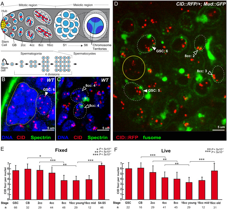
Unpaired centromeres in male germline stem cells become paired during cyst divisions. (A) Schematic overview of germ cell development in Drosophila testis. At the apical tip are the somatic hub cells (yellow), which serve as the niche for GSCs. GSCs divide asymmetrically giving rise to a GSC and a daughter cell, the GB, which undergoes four mitotic divisions by incomplete cytokinesis to produce interconnected 2-, 4-, 8-, and 16-cell spermatogonial cysts. Germ cells undergo an extended growth phase (S1 to S6) entering meiosis as primary spermatocytes. A zoom view of chromosome territories in one spermatocyte. The spectrosome (red circles) of GSCs and GB develops into a branched structure called the fusome (red) during each division. (B and C) Z-section projections of wild-type fixed testes stained for centromeres (CID, red), fusome (α-spectrin, green), and DNA (DAPI, blue). There are six centromeres in the GSC (B, arrowheads) but fewer in 8-cell cyst nuclei (C, open arrowheads), as quantified. (D) Z-section projection obtained by live imaging of a testis expressing the centromere marker CID::RFP (red) and the fusome marker MUD::GFP (green). Two GSCs (arrowheads) identified by their position next to the hub cells (yellow circle) and an 8-cell cyst (8cc), with cells linked by the fusome. Two nuclei of an 8-cell cyst (8cc, open arrowheads), with fusome GFP, are marked by dotted lines. (E and F) Graphs plot the number of CID foci for each developmental stage in the mitotic region of fixed (E) or live (F) wild-type testes. The number of analyzed cells is given below each stage. In B, C, and D, dotted lines surround germ cell nuclei. (Scale bars in B–D, 5 µm.) *P < 5 × 10⁻², **P < 5 × 10⁻³, ***P < 5 × 10⁻⁷ (two-tailed Student’s t test).

