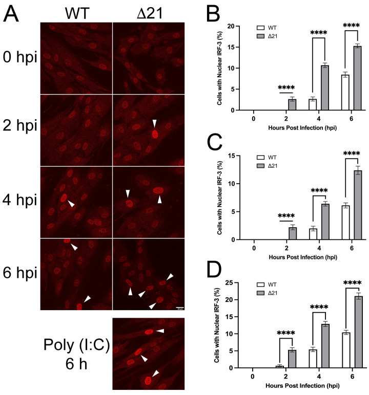
Fig 5. Examination of IRF-3 nuclear localization during early stages of infection with HSV WT and Δ21 strains.
T12 cells were infected at an MOI of 3 and after inoculation for one-hour on ice, cells were incubated in medium containing 50 μg/ml of cycloheximide to inhibit protein synthesis. Infected cells were fixed at 0, 2, 4 and 6 hpi, stained for IRF-3 and imaged by confocal microscopy. A) Representative images of HSV-1 KOS WT and Δ21 infected cells. IRF-3 localization at 0, 2, 4, and 6 hpi. Nuclear IRF-3 is indicated by the white arrowheads. Representative image of T12 cells transfected for 6 hours with poly I:C is shown as a positive control. (n = 33–37 images per timepoint and condition). Scale bar is 20μm and applies to all panels. B) Quantification of nuclear IRF-3 in cells infected with HSV-1 KOS WT and Δ21 strains (n = 33–36 images per timepoint and condition). C) Quantification of nuclear IRF-3 in cells infected with HSV-1 F WT and Δ21 strains (n = 36 images per timepoint and condition). D) Quantification of nuclear IRF-3 in cells infected with HSV-2 SD90e WT and Δ21 strains (n = 36 images per timepoint and condition). Unpaired t-tests were performed to compare the percentage of cells with nuclear IRF-3 for each timepoint in Δ21 mutants with WT virus for each strain, from three biological replicates. **** represents p ≤ 0.0001.

