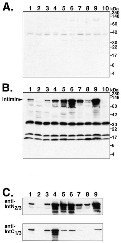
FIG. 2.
Immunoblot analysis of intimin from whole-cell lysates of different E. coli strains with antibodies raised in a goat against intimin from O157:H7 strain 86-24. The strains analyzed were as follows (lanes 1 to 8 are EHEC strains): lane 1, 86-24 (O157:H7); lane 2, 86-24eaeΔ10; lane 3, EDL933 (O157:H7); lane 4, 97-3256 (O55:H7); lane 5, 85-3007 (O111:NM); lane 6, H19 (O26:H11); lane 7, 97-3250 (O26:H11); lane 8, 95-3208 (O111:NM); lane 9, EPEC strain E2348/69 (O127:H6); lane 10, nonpathogenic E. coli K12 strain UT5600. (A) Reactivity of preimmune serum. (B) Reactivity of polyclonal serum raised against intiminO157. For blots A and B, whole-cell lysates were run on 15% polyacrylamide–SDS gels for this figure to display all of the reactive bands. Identical serum dilutions were used for the preimmune and anti-intimin antisera, and blots A and B were exposed to film simultaneously for chemiluminescent detection. (C) Reactivity of purified antibodies against the N-terminal two-thirds (top) and C-terminal third (bottom) of intimin. Bacterial lysates were run on 10% polyacrylamide–SDS gels for the blots shown here. Analysis of higher-percentage gels showed additional reactivity to a 15-kDa band in all lanes (data not shown). All immunoblots were scanned with a Hewlett-Packard ScanJet 4C scanner, and the images of the four blots were arranged for the figure and labeled with Adobe Photoshop version 5.0.2 for Macintosh.

