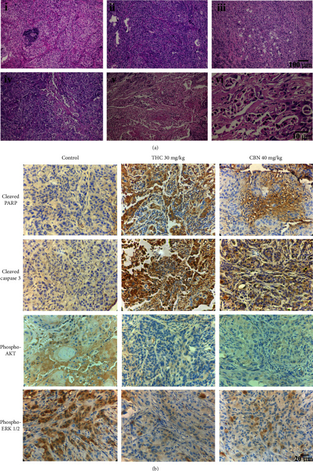
Figure 7.

Representative H&E staining and immunohistochemistry of xenograft mouse specimens. (a) H&E staining of specimens from xenograft mice treated for 21 d with (i) control, (ii) 20 mg/kg CBN, (iii) 40 mg/kg CBN, (iv) 15 mg/kg THC, (v) 30 mg/kg THC (i–v, scale bars: 100 μm), and (vi) 30 mg/kg THC (scale bar: 10 μm). (b) Immunohistochemical staining of specimens from xenograft mice treated as indicated. The specimens of xenograft mice treated with 30 mg/kg THC or 40 mg/kg CBN markedly expressed cleaved PARP and cleaved caspase 3 but scarcely expressed phosphorylated AKT and ERK1/2 compared to the specimens of the control mice (scale bars: 20 μm).
