
Figure 5.
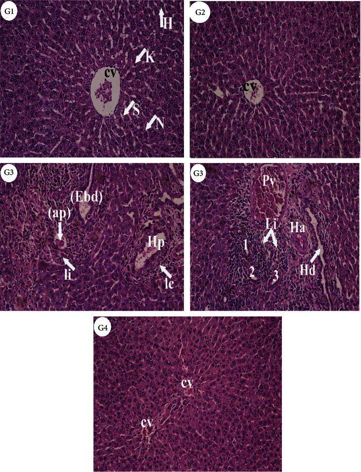
Photomicrographs of liver sections from different experimental groups stained with Hematoxylin & Eosin. Control (G1) and bromelain (G2) rats revealed the normal histological structures of normal hepatocytes (H) with nuclei (N), central vein (CV), sinusoids (S), and Kupffer cells (K) which represented monocytes-macrophage defense system. (G3), AlCl3 treated rats showed inflammation or infiltration (Ii) and enlarged bile ductular (Ebd), apoptic body (ap), branches of hepatic portal vein (Hp) surrounded by inflammatory cells (Ic). Also, leukocytic infiltration (Li) and bile ductular proliferation (1, 2, 3), branch of hepatic artery (Ha), congested portal vein (Pv) and enlarged hepatic duct (Hd) and hyperchromatic nuclei (Hn) were observed. (G4), Bromelain+AlCl3 group revealed more or less normal hepatocytes (H&E X 200).