
Fig. 6.
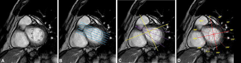
CMR planning for assessment of mitral valve. ( A ) CMR short axis view at the level of mitral valve showing anatomy of mitral valve. A1, A2, and A3 scallops in the anterior leaflet. P1, P2, and P3 scallops in the posterior leaflet. ( B ) Modified left ventricular outflow tract view. Contiguous stack of cine images perpendicular to the mitral commissures transecting the principal line of coaptation to visualize and assess all the mitral scallops A1–P1, A2–P2, and A3–P3. ( C ) Additional/alternative slices perpendicular to the coaptation plane of the valve leaflets ( yellow and/or purple lines ) ( D ) For circumferential assessment of annular plane, six left ventricular long-axis cine slices are acquired at every 30degrees. The first projection was aligned through the superior right ventricular free wall insertion into the septum, and was defined as 0° in the annular plane, followed by clockwise labeling of the long-axis slices. CMR, cardiac magnetic resonance imaging.