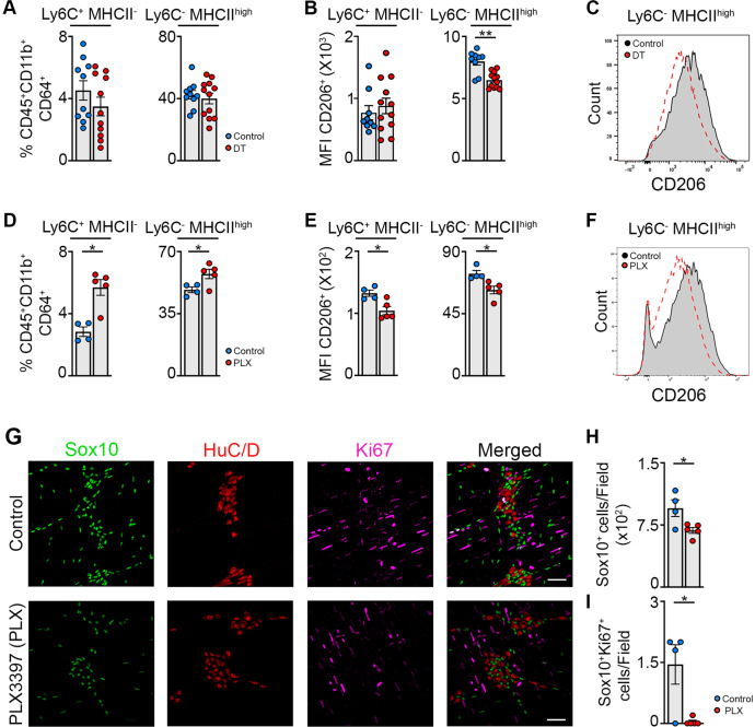
Fig. 7. EGCs are crucial for monocyte differentiation into anti-inflammatory CD206+ Mφs in part via CSF-1 in vivo.
a–c A small portion of the small intestine of PLPCreERT2 iDTR mice was exposed to saline or DT after intestinal manipulation and mice were sacrificed 72 h after the induction of muscularis inflammation. a Percentages of Ly6C+ MHCII− monocytes and Ly6C− MHCIIhi Mφs in live CD45+ CD11b+ Ly6G− CD64+ population. b Mean fluorescent intensity (MFI) of CD206 in Ly6C+ MHCII− monocytes and Ly6C− MHCIIhi Mφs. c Histogram of CD206 expression in Ly6C− MHCIIhi Mφs in control and DT exposed PLPCreERT2 iDTR mice 72 h after muscularis inflammation. d–h Wild-type mice were gavaged daily with vehicle or PLX-3397 (50 mg/kg) starting from the day of the manipulation and sacrificed 72 h after the induction of muscularis inflammation. D Percentages of Ly6C+ MHCII− monocytes and Ly6C− MHCIIhi Mφs in live CD45+ CD11b+ Ly6G− CD64+ population. e Mean fluorescent intensity (MFI) of CD206 in Ly6C+ MHCII− monocytes and Ly6C− MHCIIhi Mφs. f Histogram of CD206 expression in Ly6C− MHCIIhi Mφs in control and PLX treated mice 72 h after muscularis inflammation. g Immunofluorescent images of muscularis whole-mounts in control and PLX treated mice 72 h after muscularis inflammation stained for SOX10 (green), HuC/D (red) and Ki-67 (purple). Scale bar 25 µm. h Quantification of the number of SOX10+ cells per field in control and PLX treated mice 72 h after muscularis inflammation (average of 4–5 pictures/mouse). i Quantification of the number of Ki-67+ SOX10+ cells per field in control and PLX treated mice 72 h after muscularis inflammation (N = 4–5 pictures/mouse). a, b; d, e; h, i. T-test. *p < 0.05; **p < 0.01.

