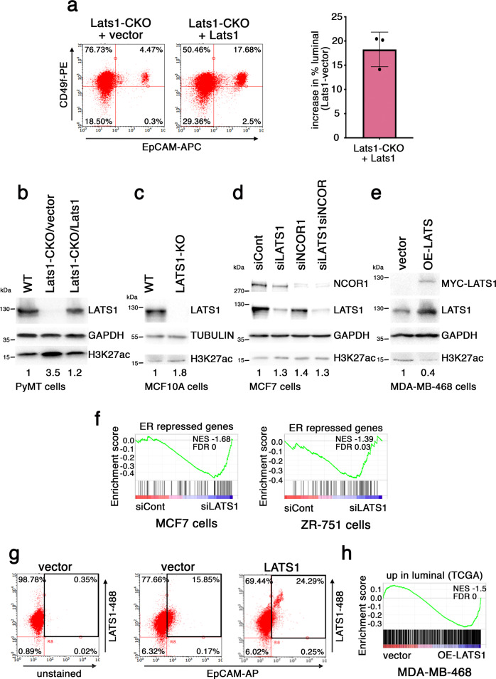
Fig. 4. LATS1 regulates phenotypic plasticity.
a MYC-tagged mouse Lats1 or vector control was stably introduced into three independent Lats1-CKO cell lines. Left: representative FACS analysis as in Fig. 3a. Right: graphical representation of the proportional increase in luminal cells in each cell line upon LATS1 overexpression, measured as in (a, left) (mean ± SD of 3 cell lines). Source data are provided as a Source Data file. b Lats1-CKO PyMT cells stably harboring vector or MYC-tagged mouse Lats1 were subjected to Western blot analysis with the indicated antibodies. A WT sample is presented in the first lane for comparison. GAPDH served as a loading control. Numbers under lanes represent relative H3K27ac band intensity, normalized to the corresponding loading control and control sample. Representative blot of five biological repeats. c MCF10A cells, either WT or with CRISPR/Cas9 deletion of LATS1 (LATS1-KO), were subjected to Western blot analysis with the indicated antibodies. Tubulin served as a loading control. Numbers are as in (b). Representative blot of two biological repeats. d MCF7 cells transiently transfected with the indicated siRNAs were subjected to Western blot analysis with the indicated antibodies. GAPDH served as a loading control. Numbers are as in (b). Representative blot of three biological repeats. e MDA-MB-468 cells harboring vector control or human MYC-LATS1, after 48 h of doxycycline induction, were subjected to Western blot analysis with the indicated antibodies. Top panel = 9E10 antibody, directed against the MYC-tag. GAPDH served as a loading control. Numbers are as in (b). Representative blot of four biological repeats. f GSEA of MCF7 (left) and ZR-751 (right) cells, transiently transfected with control siRNA (siCont) or LATS1 siRNA (siLATS1) (data from Furth et al.61), compared to an ER repressed30 gene set. g FACS analysis of MDA-MB-468 cells harboring vector control or human MYC-LATS1, after 48 h of doxycycline induction. Cells were first stained for APC-EpCAM, and then permeabilized to stain intracellular LATS1 (probed with Alexa Fluor 488 dyed secondary antibody). Black square designates the quadrant of EpCAMhigh luminal cells. h GSEA of MDA-MB-468 cells without (vector) vs. with induction of LATS1 (OE-LATS1), compared to genes upregulated in human luminal tumors relative to basal-like tumors (data from TCGA).

