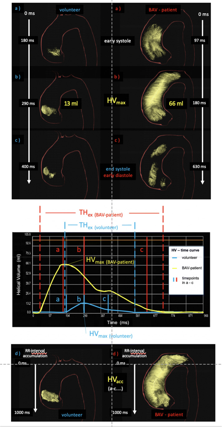
Fig. 1.
3D visualization of the evolution of a helix in the ascending aorta during the entire cardiac cycle of a healthy individual (left column) and a patient with BAV (female, 39 years old, right column). (a) At early systole, the helix occupies only the very proximal part of the ascending aorta in die healthy volunteer and a big part in the patient. (b) At mid-systole, it has grown in volume and length and reached its maximum—the maximum helical volume (HVmax). (c) At end-systole at 300 ms, most of the helix vanished. (d) The summation of all volumes that contributed to the helix during the entire cardiac cycle is the accumulated helical volume (HVacc), and its length is the accumulated helical volume length (HVLacc)

