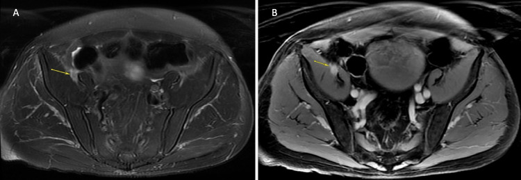
FIG. 3.
MRI of the pelvis demonstrating an enhancing metastasis to the right femoral nerve. A: Axial fat-saturated image depicting focal enlargement of the femoral nerve as it courses between the iliacus and psoas muscles (arrow). B: Axial T1-weighted postcontrast image depicting diffuse enhancement of the mass and the femoral nerve (arrow), likely secondary to melanin deposition.

