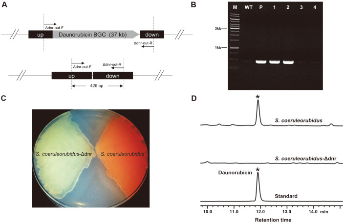
Fig. 4. Deletion of the daunorubicin BGC in S. coeruleorubidus.
(A) The schematic diagram of the double-crossover mutants. Up, the upstream homologous arm of the daunorubicin BGC; down, the downstream homologous arm of the daunorubicin BGC. The primer pair Δdnr-out-F/Δdnr-out-R was used for PCR verification. (B) PCR verification of the S. coeruleorubidus-Δdnr mutants. Lane M, DNA marker; lane WT, wild-type S. coeruleorubidus; lane P, pSUC03; lane 1-2, doublecrossover mutants (2 randomly selected colonies that secreted no light red pigments into the plate); lane 3-4, reverted wild-type colonies (2 randomly selected colonies that produced the light red compounds into the plate). (C) Color comparison of the substrate mycelium cultured for 6 days before being photographed. (D) The HPLC analysis of the fermentation broths at 120 h.

