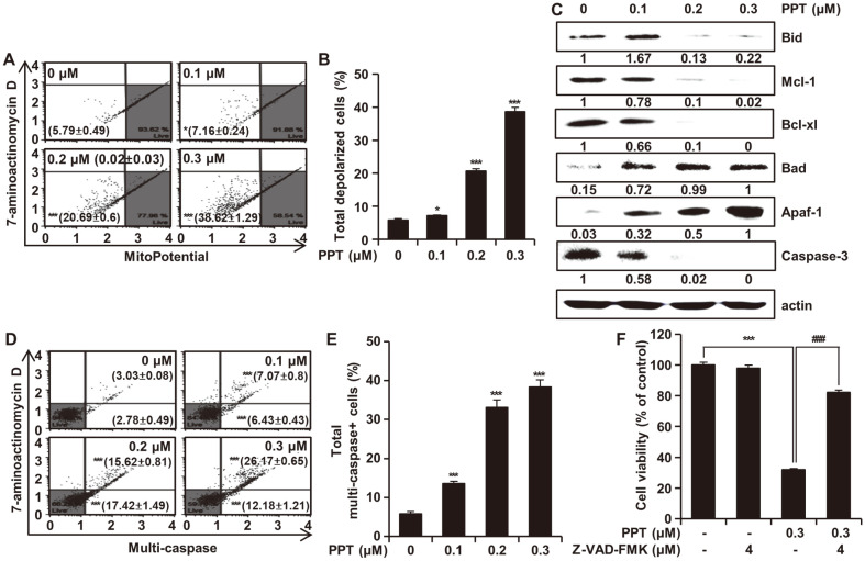
Fig. 5. Effects of PPT on Mitochondrial-Mediated Apoptosis and Caspase Activation.
(A and B) HCT116 cells were treated with PPT (0, 0.1, 0.2, and 0.3 μM) for 48 h and the extent of mitochondria membrane potential depolarization was measured using the Muse Mitopotential Kit. Combined MitoPotential dye and 7-AAD reactivity allowed classification of the cells into four groups, as follows: live cells with intact mitochondrial membrane potential (bottom right, Mitopotential+/7- AAD-), depolarized live cells (bottom left, Mitopotential-/ 7-AAD-), dead cells (top right, Mitopotential+/ 7-AAD+), and depolarized dead cells (top left, Mitopotential-/7-AAD+). (C) HCT116 cells were treated with PPT for 48h and then analyzed by Western blots with antibodies against Bad, Bid, Mcl-1, Bcl-xl, Apaf-1 and caspase-3. Actin was used as internal standard. (D and E) HCT116 cells were treated with PPT (0, 0.1, 0.2, and 0.3 μM) for 48 h and multi-caspase (caspase-1, -3, -4, -5, -6, -7, -8, and -9) was measured using the Muse MultiCaspase Kit. Cells on the bottom right side indicate caspase-positive/live cells whereas the cells on the top right side indicate caspase-positive/dead cells. (F) HCT116 cells were treated with a pan caspase inhibitor Z-VAD-FMK (4 μM) with or without PPT (0.3 μM) for 48h. Cell viability was measured by the MTT assay. All data shown represent the mean ± SD (n = 3). *p < 0.05 and ***p < 0.001 vs the control group; ###p < 0.001 vs the PPT-treated group.

