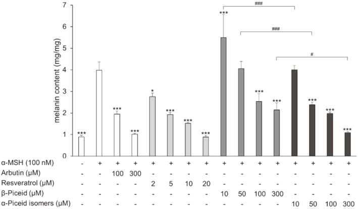
Fig. 4. Effect of drugs on cellular melanin content in α-MSH-stimulated B16F10 melanoma cells.
The cells were treated with α-MSH alone or together with arbutin, resveratrol, β-piceid, or α-piceid for 72 h and then cellular melanin content was measured. Significant differences were compared with α-MSH treated cells: *p < 0.05 and ***p < 0.001, significant differences were also observed between β-piceid and α-piceid isomers: #p < 0.05 and ###p < 0.001.

