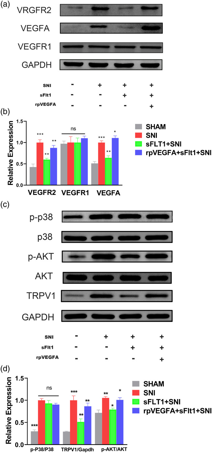
Figure 3.
Differential spinal cord VEGFA pathway expression in each group as determined by Western blot. (a–d) Molecular expression and quantification in each group. The expression of VEGFR1 (a, b) and AKT (c, d) were not different in each group, while that of p-p38 was increased after SNI surgery compared with the SHAM group, but did not different from that in the sFlt1+SNI and in rpVEGFA+sFlt1+ SNI group (c, d). The increased expression of VEGFA, VEGFR2, p-AKT, and TRPV1 was ameliorated by sFIt1 treatment (green bar, B and D), and rpVEGFA inhibited the sFIt1-induced downregulation (blue bar, B and D). The data are shown as the mean ± SEM (n = 3). The results were analyzed by One-way analysis of variance ANOVA and by multiple comparisons. ⁎ p < 0.05, ⁎⁎ p < .01, and ⁎⁎⁎ p < .001. SNI: spare nerve injury.

