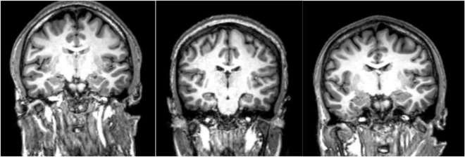
FIGURE 1.
MPRAGE images of three patients with schizophrenia. These type of high resolution T1-weighted images allow quantitation of specific gray matter and cerebrospinal fluid (CSF) containing structures which has led to the conclusion that patients with schizophrenia have overall decreased gray matter volumes and enlarged CSF spaces. Specific cortical regions are best evaluated in group analyses and/or voxel-based morphometry analyses. Comparing these three subjects, one finds reduced right versus left hippocampal volume in the second and third subjects at group level, a finding that is not necessarily confirmed by visual analysis at individual level.

