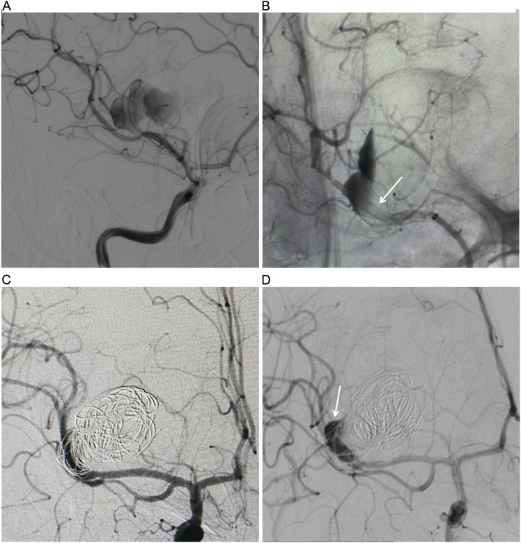
Figure 1.
(A) DSA showing the right MCA M2 dissecting aneurysm. (B) DSA in working position view immediately after flow diversion shows diminished filling of the right MCA dissecting aneurysms. The expanded Pipeline flow diversion device in the MCA M1 to M2 segment (arrow). (C) DSA in AP view immediately after PED-assisted coil embolization shows diminished filling of the aneurysm. (D) Six-month DSA follow-up shows persistence after flow diversion at the aneurysm nack (arrow).

