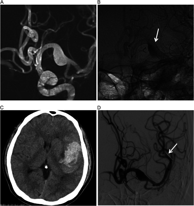
Figure 4.
(A) DSA fluoroscopic roadmap image showing a right MCA M1 dissecting aneurysm. (B) DSA in working position views immediately after flow diversion showing diminished filling of aneurysms (arrow). (C) CT showing hemorrhage in the left temporal lobe. (D) The micro delivery wire damaged the small distal artery during the release of the stent (arrow).

