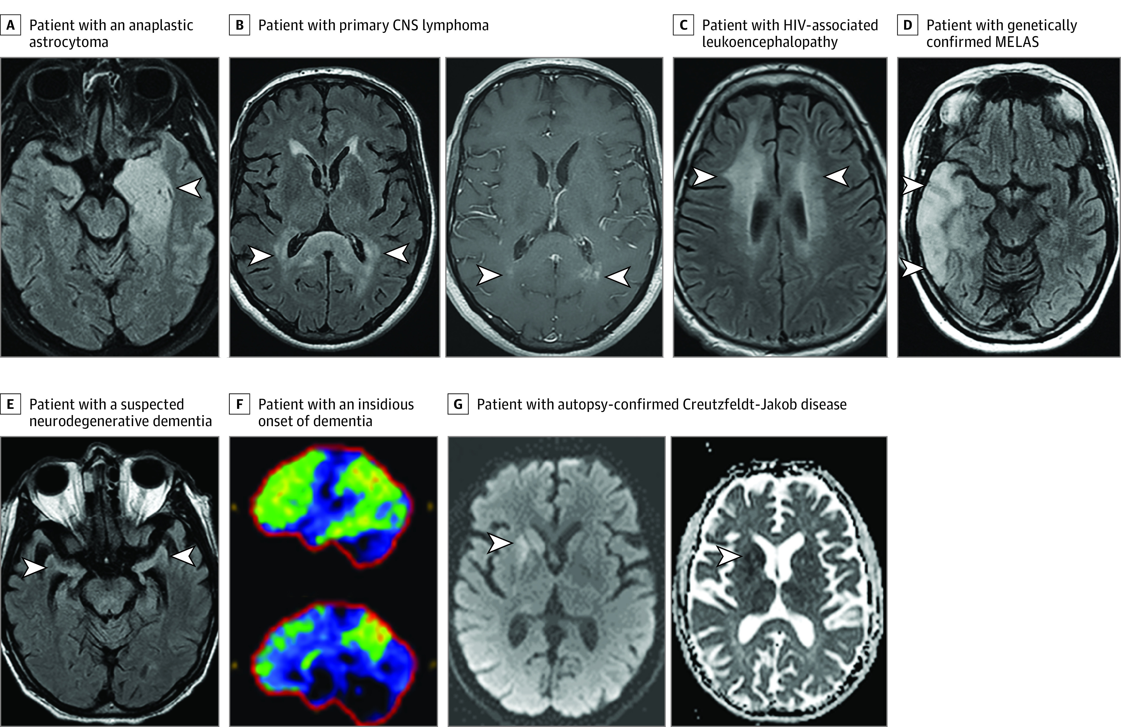
Figure. Imaging Examples of Patients Who Were Initially Thought to Have Autoimmune Encephalitis but Later Had an Alternative Diagnosis Made.

A T2-weighted axial fluid-attenuated inversion recovery (T2-FLAIR) image reveals a left mesial temporal lobe T2-hyperintensity and swelling (A, arrowhead) in a patient with an anaplastic astrocytoma. Note in retrospect the fullness/enlargement of the affected region, possibly suggesting some mass effect. Axial T2-FLAIR image reveals bilateral splenium T2-hyperintensity (B, left panel, arrowheads) with multifocal punctate enhancement (B, right panel, arrowheads) in a patient with primary central nervous system (CNS) lymphoma. An axial T2-FLAIR image reveals bilateral confluent T2-hyperintensity in the subcortical white matter (C, arrowheads) in a patient with HIV-associated leukoencephalopathy. Axial T2-FLAIR image reveals right temporal cortical swelling and T2-hyperintensity (D, arrowheads) in a patient with genetically confirmed mitochondrial encephalomyopathy, lactic acidosis, and stroke-like episodes (MELAS). An axial T2-FLAIR image shows disproportionate bilateral hippocampal atrophy (E, arrowheads) in a patient with a suspected neurodegenerative dementia with features potentially consistent with mixed Alzheimer disease and dementia with Lewy bodies. 18F-Fluorodeoxyglucose positron emission tomography reveals reduced uptake of glucose (normal, dark blue/black; mildly reduced, green; moderately reduced, yellow; severely reduced, red) in the frontotemporoparietal region, precuneus and posterior cingulate (F) most suspicious for underlying Alzheimer disease in a patient with an insidious onset of dementia and elevated cerebrospinal fluid phospho-Tau and low cerebrospinal fluid amyloid-β 42 also suggestive of this diagnosis. Axial diffusion weighted hyperintensity (G, left panel) and apparent diffusion coefficient hypointensity (G, right panel) consistent with restricted diffusion in the right caudate and putamen in a patient in whom autopsy later confirmed Creutzfeldt-Jakob disease.
