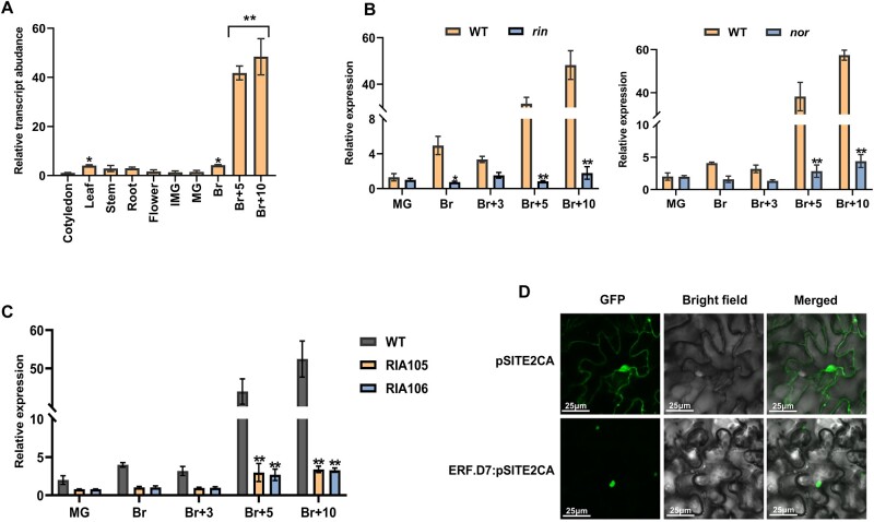
Figure 1.
Transcript profiling of SlERF.D7 (Solyc03g118190) and its subcellular localization. A, Expression of SlERF.D7 in various tissues, including cotyledon, roots, stems, leaves, flowers, and fruit at different developmental stages: immature green (IMG), MG, Br, 5-day after Br (Br+5), and 10-day after Br (Br+10) in WT (Pusa Ruby). Values are means ± sd of three independent replicates. Asterisks indicate the statistical significance using ANOVA: *, 0.01 < P-value < 0.05; **, 0.001 < P-value < 0.01. B and C, Relative expression levels (in fold change) of SlERF.D7 in WT (Ailsa Craig), ripening-inhibitor (rin; accession no. LA1795, in the unknown background) mutant and nor mutant (in Ailsa Craig background), and 3S:RIN knockdown lines (RIA105 and RIA106; in Pusa Ruby background) at various stages of fruit ripening. Expression profiles were studied at different stages of fruit ripening by employing the RT-qPCR technique. The mRNA levels of SlERF.D7 at the MG stage in WT were used as the reference for all stages. Values are means ± sd of three independent replicates. Asterisks indicate the statistical significance using Student’s t test: *, 0.01 < P-value < 0.05; **, 0.001 < P-value < 0.01. D, Subcellular localization of SlERF-D7 in the nucleus of N. benthamiana epidermal cells.

