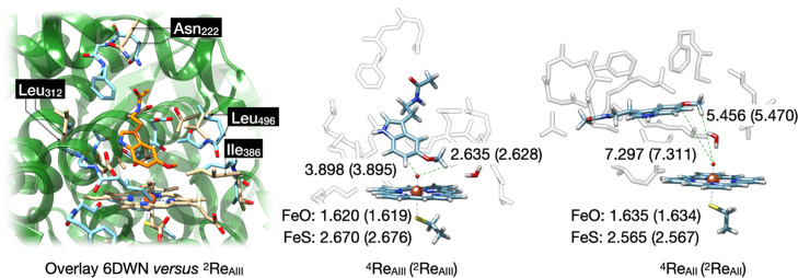
Figure 3.
(Left) Overlay of the active site of chain A of the 6DWN pdb file (green ribbons and beige atoms) with 2ReAIII (light blue). The substrate is shown in orange. (Right) QM cluster optimized geometries of the CYP1A1 reactant complexes with melatonin bound as obtained in the quartet and doublet (data in parenthesis) spin states. Bond lengths are in angstroms.

