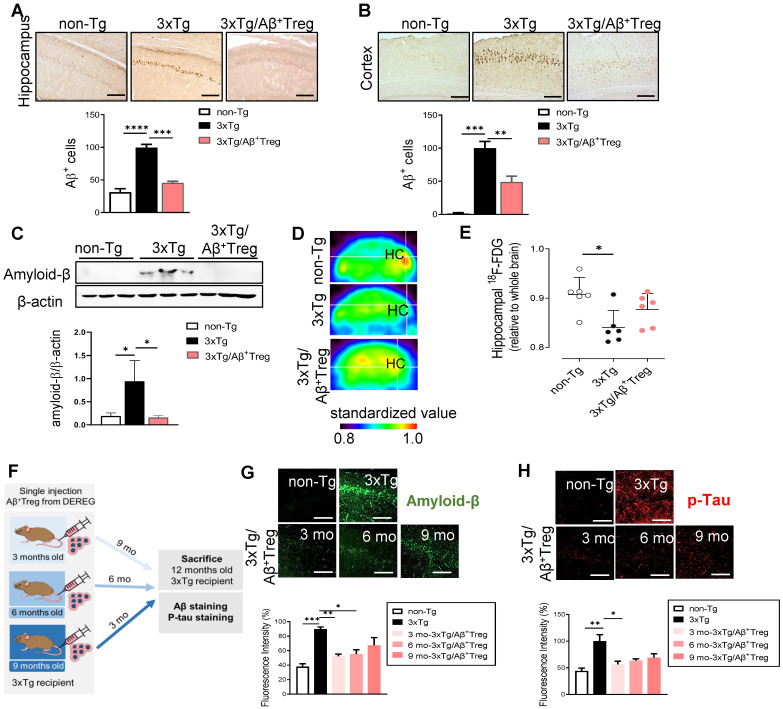
Figure 3.
Aβ-stimulated Treg transfer reduces both Aβ deposition and tau phosphorylation in AD model mice. After behavioral tests were performed, mouse brains were prepared for immunohistochemistry. (A) Aβ peptides were detected in the hippocampus and (B) cortex of 3xTg-AD mice. The scale bar represents 500 μm. (C) Proteins were extracted from the hippocampus, and the levels of Aβ were measured using western blotting. (D and E) Glucose metabolism was measured using the level of hippocampal 18F-FDG determined with a PET scan. (F) Experimental scheme for assessing Aβ+ Treg properties in different disease stages in 3xTg-AD mice. (G) Mouse brain sections were labeled for Aβ (scale bar represents 100 μm), and the staining intensity was measured. (H) Phosphorylated tau peptides were also detected in the hippocampus (scale bar represents 100 μm), and the staining intensity was measured. Data are presented as the mean ± SEM. n = 5-6; *P < 0.05, **P < 0.01, *** P < 0.001, **** P < 0.0001.

