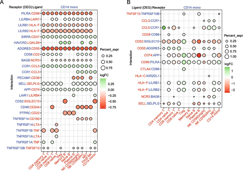
Fig. 5.
Cell–cell communication analysis. Dot plot of selected receptor/ligand pair (A) and ligand/receptor (B) interactions between CD14 + monocytes and other cell components in the COVID-19 patient group. Gene expression is indicated as log2(FC) for differentially expressed genes (FDR < 0.05), which, in both cases (A and B), are the molecules presented on the left. The percentage expression of the differentially expressed genes in each cell type is indicated by the circle size. Molecules shown in blue are those expressed in CD14 + monocytes. Molecules expressed in the immune cell partner are shown in red

