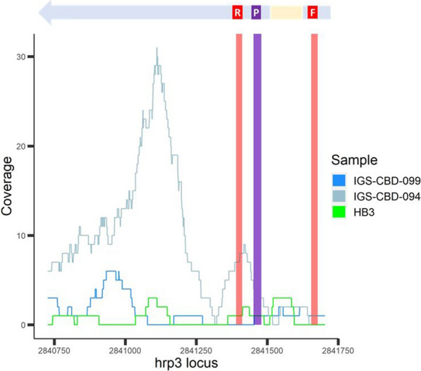
Fig. 4.

Coverage of hrp3 among select validated samples. Coverage plot of hrp3 locus of validated samples IGS-CBD-099, IGS-CBD-094 and strain HB3 (known hrp3 deletion genotype) with hrp3 schematic representation above the plot. Red areas denote the primer binding sites, and purple area denotes the probe binding site of the hrp2/3-specific qPCR assay. GC3 assigned IGS-CBD-099 as hrp3-absent; however the 5′ end of the gene was amplified by qPCR. As a comparator, IGS-CBD-094 is a similar sample (apparent deletion at qPCR primer binding site in exon 1) that GC3 assigned as hrp3-present. HB3 is a laboratory reference strain known to be missing the hrp3 locus. It is important to note some position with non-zero coverage in HB3, which suggests that non-orthologous reads from HB3 map the hrp3 locus of Pf3D7
