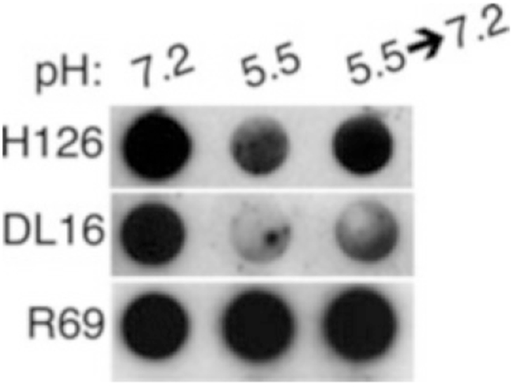
Fig. 3.

Dot blot illustrating partial reversibility of conformational change in HSV-1 gB. HSV-1 KOS virions were treated with medium buffered to pH 7.2 or 5.5. For the indicated samples, pH was neutralized back to 7.2 for 10 min at 37 °C. Membranes were probed at neutral pH with the indicated antibodies, followed by horseradish peroxidase-conjugated secondary antibody (Reproduced from ref. 9 with permission from the American Society for Microbiology)
