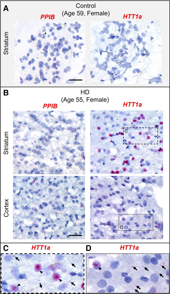
Figure 5.
Hs HTT1a forms clusters in post-mortem HD brain and are detectable in the cytoplasm as foci Chromogenic RNAscope assay was performed in healthy control and HD post-mortem human brains and counterstained with hematoxylin. (A) PPIB and HTT1a mRNA in post-mortem control striatum. Scale bar, 20 µm. (B) PPIB and HTT1a mRNA in post-mortem HD striatum (top) and cortex (bottom). Scale bar, 20 µm. (C, D) Insets of boxed regions in panel (B) showing HTT1a mRNA in the striatum (C) and cortex (D). Arrowheads indicate HTT1a clusters, and arrows indicate cytoplasmic HTT1a foci.

