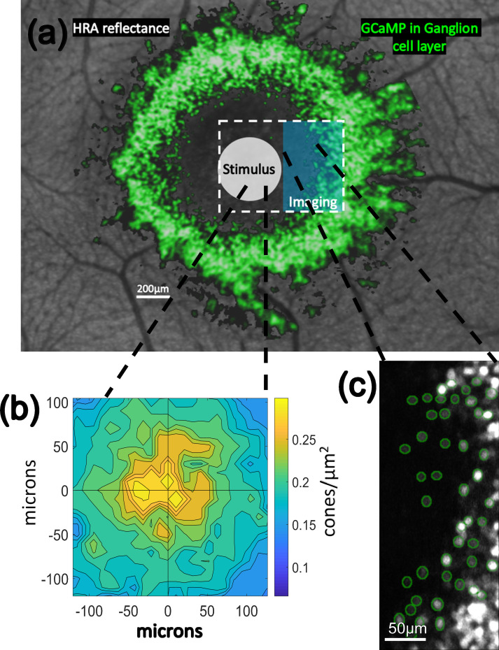
Fig 1. Stimulus presentation and recording paradigm.
In (a) the general stimulation paradigm is shown. Background is an image of the retina of M3 using the blue reflectance imaging modality of a Heidelberg Spectralis instrument. In false color green are GCaMP-expressing cells from M3 imaged using the blue autofluorescence modality of the same Spectralis instrument. When using the AOSLO instrument, videos are captured within a rectangular field of view (white dashed line) while stimuli (white circle) such as cone-isolating flicker or drifting gratings (which were rectangular in shape and covered approximately the same area as the white circle) are presented to and centered on the centermost foveal cone photoreceptors. A 488 nm imaging laser in the AOSLO (blue rectangle) excites GCaMP-mediated fluorescent responses of ganglion cells surrounding the foveal slope. (b) shows the recorded cone densities at the fovea of M3 and (c) shows the ganglion cells segmented (green) at the innermost edge of the foveal slope in M3.

