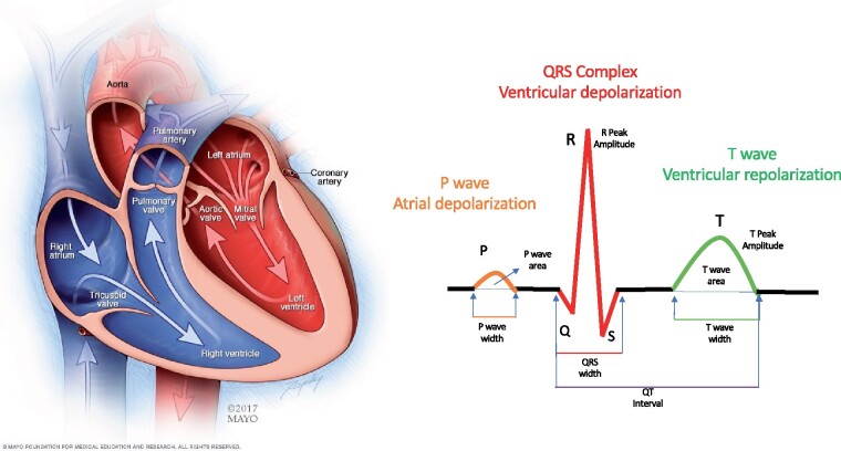
Figure 1.
Cardiac anatomy and the electrocardiogram signal. The heart has four chambers. The upper chambers (the atria) are activated by the signal reflected in the electrocardiogram as the P-wave. The lower chambers (the ventricles) are rapidly activated resulting in the QRS complex; the relaxation of the ventricles (repolarization) is represented by the smoother T-wave. A number of human-selected features, such as the peak amplitude of the various waves, the areas and widths of the different waves, deviation from baseline, and other morphological characteristics have a known biological mechanism and associations with specific pathologies.

