Fig. 2.
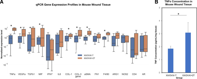
Murine wound tissue analysis. A, qPCR gene expression profiles of mouse wound tissue. Relative expression levels of cytokine (Tumor Necrosis Factor alfa, Vascular Endothelial Growth Factor A, Transforming Growth Factor beta-1, Macrophage Migration Inhibitory Factor, Interferon gamma, Interleukin 6) extracellular matrix (Collagen 1, Col 3, Smooth Muscle alpha-actin, Fibronectin-1), cell phenotype (F4/80, Arginase-1, Nitric Oxide Synthase-2, FOXP3, CD4), and androgen receptor (AR)-related genes (n = 12; P < 0.05). B, ELISA of TNFα in mouse skin wound tissue (n = 6; P < 0.05). *indicates significance of P < 0.05.

