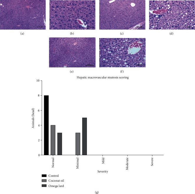
Figure 3.

Hepatic macrovesicular steatosis. The histopathological staining of liver tissues showing hepatic macrovesicular steatosis in the control group at 10x (a) and 40x (b), coconut oil-fed group at 10x (c) and 40x (d), omega lard-fed group at 10x (e) and 40x (f) and the hepatic macrovesicular steatosis scores (g). Increased incidences of hepatic macrovesicular steatosis were observed in both the coconut oil- and omega lard-fed groups compared to the control group. The hepatic macrovesicular steatosis scores were consistent, showing a slightly increased incidence of steatosis in both the coconut oil- and omega lard-fed groups. No significant difference between the coconut oil- and omega lard-fed groups was observed (chi-square test, P < 0.05). The number of mice is presented on the y-axis and the severity of hepatic macrovesicular steatosis on the x-axis. Liver tissues were harvested from all animals at the end of the experiment (four weeks). The number of animals was 8, 7, and 8 in the control, coconut oil-fed, and omega lard-fed groups, respectively.
