FIGURE 3.

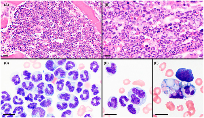
Bone marrow core biopsy (A and B) and aspirate (C‐E) specimens showing high cellularity and myeloid hyperplasia. Maturation of the myeloid lineage was complete through segmented neutrophils with prominent band neutrophils in many regions (B and C). Few giant neutrophils (D) and rare neutrophil phagocytosis (E) were present. H&E stain of B5‐fixed tissue (A and B) and Wright stain (C‐E); magnification bars = 20 μm (A), 10 μm (B‐E)
